
Fitxer:6nb6 prefusion 6m3w postfusion spike.png
Fitxer original (768 × 1.536 píxels, mida del fitxer: 382 Ko, tipus MIME: image/png)
| Aquest fitxer i la informació mostrada a continuació provenen del dipòsit multimèdia lliure Wikimedia Commons. |
Resum
| Descripció |
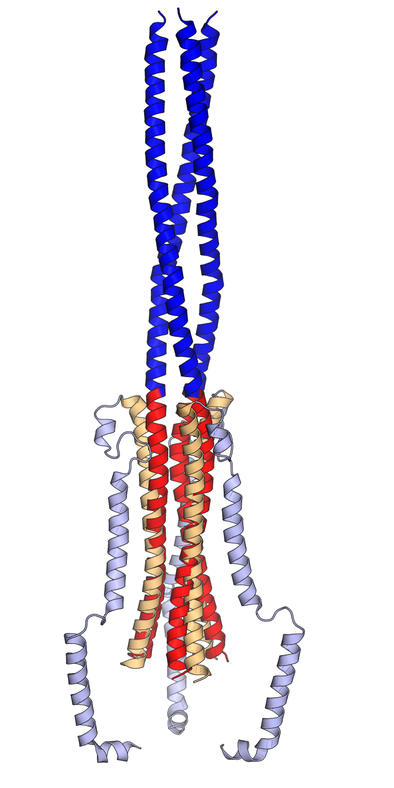
Comparison of the pre-fusion (orange, light blue) and post-fusion (red, dark blue) conformations of the SARS-CoV spike protein trimer. In the pre-fusion conformation, the central helix (orange) and heptad repeat 1 (HR1, light blue) are folded back on each other in an antiparallel orientation. In the post-fusion conformation, the central helix (red) and the HR1 sequence (dark blue) reorganize to form an extended trimeric coiled coil. The viral membrane is at the bottom and the host cell membrane at the top. Only key portions of the S2 subunit are shown. Rendered using PyMol from cryo-electron microscopy structures PDB: 6NB6 (pre-fusion) and PDB: 6M3W (post-fusion) superposed using the central helix sequences, inspired by Figs 1 and 2 from Fan 2020. 6NB6: Unexpected Receptor Functional Mimicry Elucidates Activation of Coronavirus Fusion. Walls, A.C., Xiong, X., Park, Y.J., Tortorici, M.A., Snijder, J., Quispe, J., Cameroni, E., Gopal, R., Dai, M., Lanzavecchia, A., Zambon, M., Rey, F.A., Corti, D., Veesler, D. (2019) Cell 176: 1026-1039.e15 PubMed: 30712865 DOI: 10.1016/j.cell.2018.12.028 6M3W: Cryo-EM analysis of the post-fusion structure of the SARS-CoV spike glycoprotein. Fan, X., Cao, D., Kong, L., Zhang, X. (2020) Nat Commun 11: 3618-3618 PubMed: 32681106 DOI: 10.1038/s41467-020-17371-6 |
| Data | |
| Font | Treball propi |
| Autor | Opabinia regalis |
Llicència
- Sou lliure de:
- compartir – copiar, distribuir i comunicar públicament l'obra
- adaptar – fer-ne obres derivades
- Amb les condicions següents:
- reconeixement – Heu de donar la informació adequada sobre l'autor, proporcionar un enllaç a la llicència i indicar si s'han realitzat canvis. Podeu fer-ho amb qualsevol mitjà raonable, però de cap manera no suggereixi que l'autor us dóna suport o aprova l'ús que en feu.
- compartir igual – Si modifiqueu, transformeu, o generareu amb el material, haureu de distribuir les vostres contribucions sota una llicència similar o una de compatible com l'original

|
S'autoritza la còpia, la distribució i la modificació d'aquest document sota els termes de la llicència de documentació lliure GNU versió 1.2 o qualsevol altra versió posterior que publiqui la Free Software Foundation; sense seccions invariants, ni textos de portada, ni textos de contraportada. S'inclou una còpia d'aquesta llicència en la secció titulada GNU Free Documentation License. |
Historial del fitxer
Cliqueu una data/hora per veure el fitxer tal com era aleshores.
| Data/hora | Miniatura | Dimensions | Usuari/a | Comentari | |
|---|---|---|---|---|---|
| actual | 09:02, 13 set 2021 | | 768 × 1.536 (382 Ko) | Opabinia regalis | {{Information |Description=Comparison of the pre-fusion (orange, light blue) and post-fusion (red, dark blue) conformations of the SARS-CoV spike protein trimer. In the pre-fusion conformation, the central helix (orange) and heptad repeat 1 (HR1, light blue) are folded back on each other in an antiparallel orientation. In the post-fusion conformation, the central helix (red) and the HR1 sequence (dark blue) reorganize to form an extended trimeric coiled coil. The viral membrane is at the bott... |
Ús del fitxer
La pàgina següent utilitza aquest fitxer:
Ús global del fitxer
Utilització d'aquest fitxer en altres wikis:
- Utilització a ar.wikipedia.org
- Utilització a de.wikipedia.org
- Utilització a en.wikipedia.org
- Utilització a es.wikipedia.org