Vés al contingut

Fitxer:Free-living amebic infections.png

El contingut de la pàgina no s'admet en altres llengües.
De la Viquipèdia, l'enciclopèdia lliure

Fitxer original(1.365 × 1.170 píxels, mida del fitxer: 715 Ko, tipus MIME: image/png)

Descripció a Commons
Aquesta imatge (de tipus biology) s'hauria de tornar a crear utilitzant gràfics vectorials com ara un fitxer SVG. Això té diversos avantatges; en trobareu més informació a Commons:Media for cleanup. Si ja disposeu d'una versió d'aquesta imatge en format SVG, us preguem que la pengeu; després, reemplaceu aquesta plantilla amb la plantilla {{Vector version available|nom nou de la imatge.svg}} en aquesta imatge.

Resum

Descripció
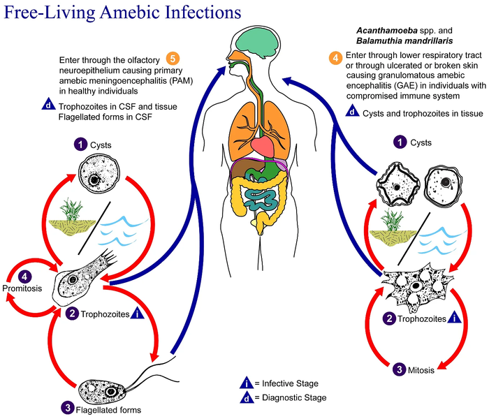
English: This is an illustration of the life cycle of the parasitic agents responsible for causing “free-living” amebic infections.

For a complete description of the life cycle of these parasites, select the link below the image or paste the following address in your address bar: http://www.dpd.cdc.gov/dpdx/HTML/FreeLivingAmebic.htm Free-living amebae belonging to the genera Acanthamoeba, Balamuthia, and Naegleria are important causes of disease in humans and animals. Naegleria fowleri produces an acute, and usually lethal, central nervous system (CNS) disease called primary amebic meningoencephalitis (PAM). N. fowleri has three stages, cysts (1) , trophozoites (2) , and flagellated forms (3) , in its life cycle. The trophozoites replicate by promitosis (nuclear membrane remains intact) (4) . Naegleria fowleri is found in fresh water, soil, thermal discharges of power plants, heated swimming pools, hydrotherapy and medicinal pools, aquariums, and sewage. Trophozoites can turn into temporary flagellated forms which usually revert back to the trophozoite stage. Trophozoites infect humans or animals by entering the olfactory neuroepithelium (5) and reaching the brain. N. fowleri trophozoites are found in cerebrospinal fluid (CSF) and tissue, while flagellated forms are found in CSF.

Acanthamoeba spp. and Balamuthia mandrillaris are opportunistic free-living amebae capable of causing granulomatous amebic encephalitis (GAE) in individuals with compromised immune systems. Acanthamoeba spp. have been found in soil; fresh, brackish, and sea water; sewage; swimming pools; contact lens equipment; medicinal pools; dental treatment units; dialysis machines; heating, ventilating, and air conditioning systems; mammalian cell cultures; vegetables; human nostrils and throats; and human and animal brain, skin, and lung tissues. B. mandrillaris however, has not been isolated from the environment but has been isolated from autopsy specimens of infected humans and animals. Unlike N. fowleri, Acanthamoeba and Balamuthia have only two stages, cysts (1) and trophozoites (2) , in their life cycle. No flagellated stage exists as part of the life cycle. The trophozoites replicate by mitosis (nuclear membrane does not remain intact) (3) . The trophozoites are the infective forms and are believed to gain entry into the body through the lower respiratory tract, ulcerated or broken skin and invade the central nervous system by hematogenous dissemination (4). Acanthamoeba spp. and Balamuthia mandrillaris cysts and trophozoites are found in tissue.
Data
Font

This media comes from the Centers for Disease Control and Prevention's Public Health Image Library (PHIL), with identification number #3412.

Note: Not all PHIL images are public domain; be sure to check copyright status and credit authors and content providers.


العربية | Deutsch | English | македонски | slovenščina | +/−

Autor
  • Photo Credit:
  • Content Providers(s): CDC/Alexander J. da Silva, PhD/Melanie Moser
Permís
(Com reutilitzar aquest fitxer)
English: None - This image is in the public domain and thus free of any copyright restrictions. As a matter of courtesy we request that the content provider be credited and notified in any public or private usage of this image.
Altres versions

Llicència

Public domain
This image is a work of the Centers for Disease Control and Prevention, part of the United States Department of Health and Human Services, taken or made as part of an employee's official duties. As a work of the U.S. federal government, the image is in the public domain.

Registre original de càrregues

Transferred from en.wikipedia to Commons by Optigan13.

La pàgina de descripció original era aquí. Els noms d'usuari a continuació es refereixen a en.wikipedia.
  • 2006-05-04 01:09 Keenan Pepper 518×435×8 (31658 bytes) Free-living_amebic_infections.gif, converted to [[PNG]] by [[netpbm]].

Llegendes

Afegeix una explicació d'una línia del que representa aquest fitxer

Elements representats en aquest fitxer

representa l'entitat

Historial del fitxer

Cliqueu una data/hora per veure el fitxer tal com era aleshores.

Data/horaMiniaturaDimensionsUsuari/aComentari
actual11:24, 2 feb 2023Miniatura per a la versió del 11:24, 2 feb 20231.365 × 1.170 (715 Ko)Materialscientisthttps://answersingenesis.org/biology/microbiology/the-genesis-of-brain-eating-amoeba/
08:30, 20 jul 2008Miniatura per a la versió del 08:30, 20 jul 2008518 × 435 (31 Ko)Optigan13{{Information |Description={{en|This is an illustration of the life cycle of the parasitic agents responsible for causing “free-living” amebic infections. For a complete description of the life cycle of these parasites, select the link below the image

No hi ha pàgines que utilitzin aquest fitxer.

Ús global del fitxer

Utilització d'aquest fitxer en altres wikis:

Metadades